
Tracing cell development with barcodes
There are different views about how blood cells develop – but they are almost exclusively based on experiments that reflect snapshots. The classic model is that different linages of blood cells spread out like the branches of a tree. The trunk is composed of stem cells and the boughs are the different progenitor cells that can give rise to different cell types. These branch out into the specialist blood cells, the red blood cells, platelets and various white blood cells that are part of the immune system. In recent years, however, increasing doubt has been cast on this model.
Scientists at the German Cancer Research Center (DKFZ) and the Max Delbrück Center for Molecular Medicine (MDC) have now presented a new technique in the journal Nature that enables us to understand this process dynamically. Using a “random generator,” research teams led by Hans-Reimer Rodewald and Thomas Höfer (DFKZ) labeled hematopoietic stem cells with genetic barcodes. This allowed them to clearly identify their descendants and track which cell types emerged from which stem cell. The technique offers new insights into the development of various tissues and carcinogenesis.
Which stem cells do blood cells come from?
“Our bar codes can be tissue-specifically induced directly in the genetic material of mice –without affecting their physiological development,” says Rodewald. The Cre/loxP system provides the basis for this approach. It can be used to recombine or remove specially marked sections of DNA. To do this, his team bred mice that carry the basic elements of the barcodes in their genomes. At a selected location where no hereditary factors are encoded, there are nine DNA snippets from a plant, the thale cress (Arabidopsis thaliana). These elements are flanked by a total of ten genetic interfaces called loxP. When the molecular scissors that match them, called Cre, are activated, random code elements in the animals’ blood stem cells are recombined or resected.
“This genetic random generator can generate up to 1.8 million different genetic barcodes, and we can identify the codes that only occur once in an experiment,” says Höfer. “The mice do the rest of the work,” says Rodewald. When blood stem cells marked in this way divide and mature, the barcodes are preserved.
In collaboration with Sascha Sauer’s team at the Scientific Genomics Platform, supported by the Berlin Institute of Medical systems biology (BIMSB) and the Berlin Institute of Health (BIH), the scientists at the MDC have conducted extensive barcode analysis to track which stem cells a particular blood cell comes from.
Fully readable barcodes were crucial
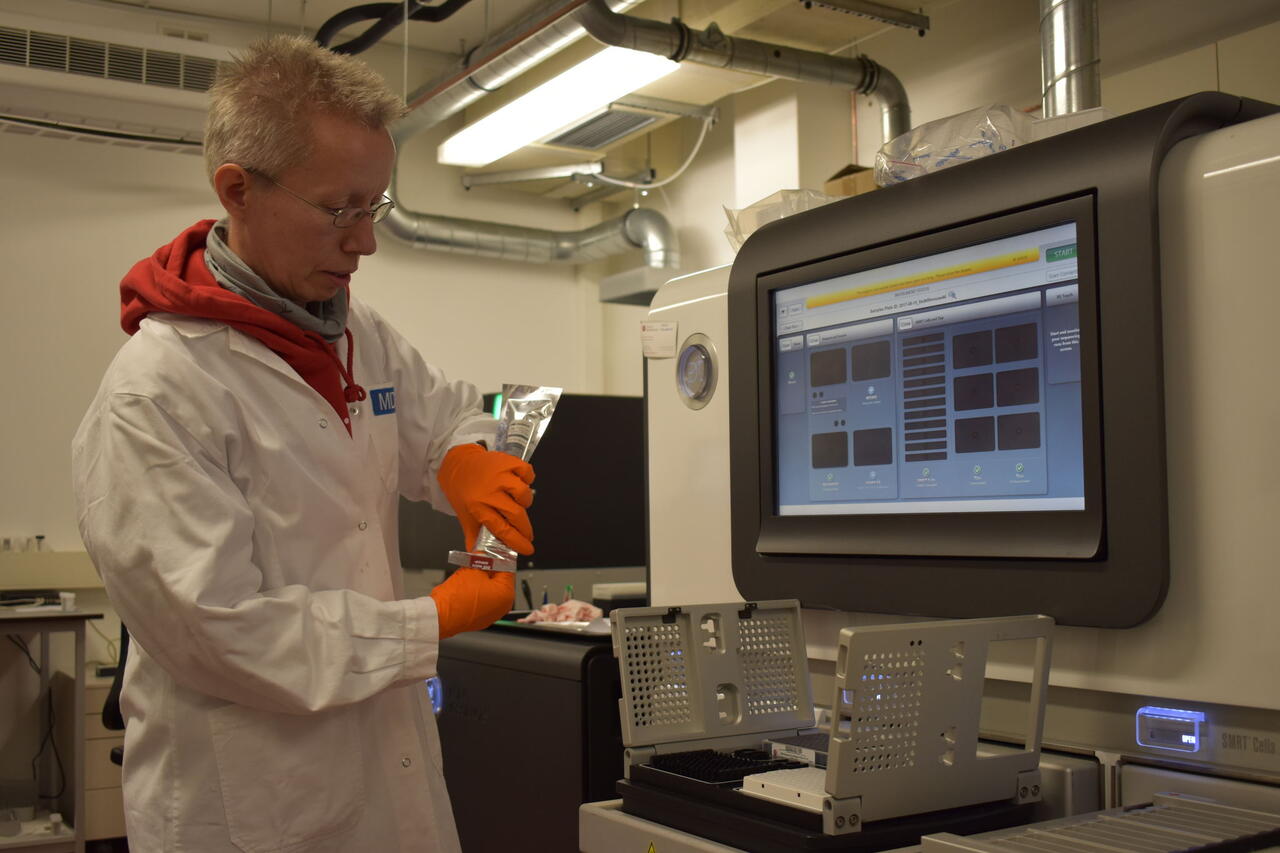
“The bar codes should not be shredded or only partially readable. Therefore it was crucial that the PacBio sequencer can directly analyze relatively long pieces of DNA with a thousand nucleotides,” says Sauer. In 2011, German Chancellor Angela Merkel pushed the start button for this sequencer’s predecessor at the Scientific Genomics Platform. Since then, the scientists have gained extensive experience with using it and can implement projects quickly and efficiently. Engineer Claudia Quedenau is continuously improving the biochemical preparation of samples and increasing throughput with more automation. A successor device is already in use.
“For the current paper, we established bioinformatics methods that efficiently analyze the bar codes and trace the development of the blood cell types,” says Sauer. This made it possible to directly observe individual bone marrow stem cells during a dynamic process. Through the small differences in the barcodes of different cells, the scientists can practically watch how the blood cell family tree branches out.
These analyses have shown that two big branches develop from the mouse blood stem cells. The T and B cells of the immune system develop in one branch. Red blood cells and various other white blood cells, such as granulocytes and monocytes, develop in the other. All these cell types can emerge from a single stem cell. “Our findings show that the classic model of hierarchical development from pluripotent stem cells applies to blood formation,” stresses Rodewald.
Collaboration with the German Cancer Research Center will continue
The Heidelberg team’s system can be used for more than investigating the development of blood cells. The strategy can in principle be applied to any tissue. In the future, this approach could be used to trace the origins of leukemia or fundamental processes of cell development in vivo in animal models. “The cooperation between the MDC and the DKFZ will continue,” says Sauer. “For me, the project is also an example of what the PacBio sequencer can do.”
The device is now capable of reading up to 20,000 nucleotides in one go. This makes it possible, for instance, to detect and quantify different isoforms of the RNA of the same gene in order to investigate the regulation of messenger RNA or biological questions about inflammation processes. “The technology is also often used to decode microbial genomes for which there is no reference sequence and to investigate viral infection processes in elephants, koala or polar bears, which the Institute for Zoo and Wildlife Research (IZW) in Berlin is interested in,” says Sauer. “There really are a lot of diverse applications.”
Further information
- Pei, W. et al (2017): “Polylox barcoding reveals haematopoietic stem cell fates realized in vivo.” Nature. doi:10.1038/nature23653
- Scientific Genomics Platform at the BIMSB and BIH
- Hans-Reimer Rodewald’s Lab at the DKFZ


